
 대표어
대표어


권호기사보기
| 기사명 | 저자명 | 페이지 | 원문 | 기사목차 |
|---|
결과 내 검색
동의어 포함
Title Page
Contents
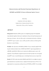
ABSTRACT 5
Introduction 7
Materials and methods 10
1. Cell lines and samples 10
2. Collection of mouse embryos 10
3. Semiquantitative reverse transcription (RT)-PCR 10
4. Overexpression of HOXB5 and HOXC10 in SNU638 gastric cancer cell line 12
5. Small interfering RNA transfection 12
6. Western blot analysis 13
7. Wound migration assay 13
8. Matrigel invasion assay 14
9. Statistical analysis 14
Results 15
1. HOX genes expression in BLBC/b mice 15
2. HOX genes expression in gastric carcinoma tissue 15
3. Stable overexpression of HOXB5 and HOXC10 in SNU 638 cell line. 15
4. Knockdown of HOXB5 and HOXC10 in TMK-1 cell line 15
5. Effect of HOXB5 and HOXC10 on proliferation of gastric cancer cells 16
6. Effect of HOXB5 and HOXC10 on migration and invasion in gastric cancer cell lines. 16
7. Involvement of MAPKs during induced and knockdown HOXC10 expression respectively. 16
8. Induction of migration and invasion by HOXB5 overexpression via Wnt/β-catenin signaling pathway. 17
Discussion 18
Reference 31
초록 35
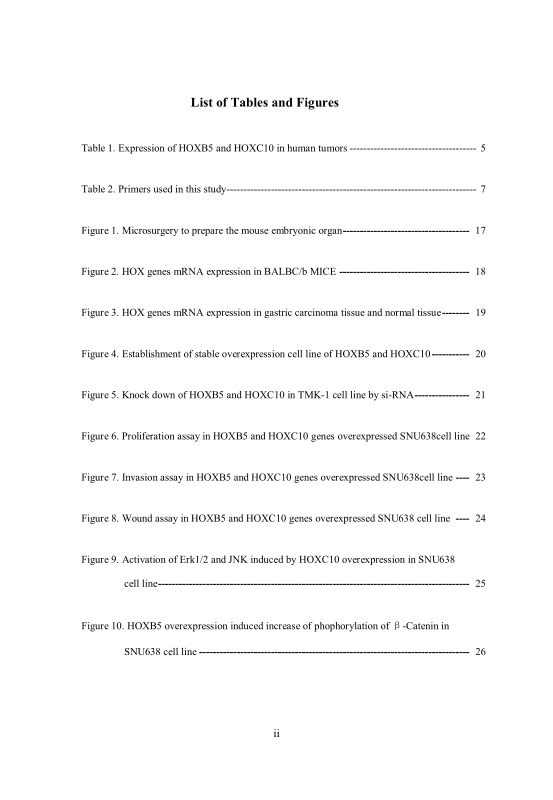
Figure 1. Microsurgery to prepare the mouse embryonic tissues. 21
Figure 2. HOX genes expression in BALB/c mice. HOXB5, HOXB13, HOXC9 and HOXC10 genes were highly expressed in embryo mouse tissues (A) compared with adult mouse tissues (B). 22
Figure 3. The genes expression in gastric carcinoma tissue and adjacent normal tissue. HOXB5 and HOXC10 genes were highly expressed in carcinoma tissues compared with noncancerous mucosa tissues (A). 23
Figure 4. HOXB5 and HOXC10 expressed in SNU638 cell line. HOXB5 and HOXC10 and pcDNA6 were transfected to SNU638 gastric cancer cell lines. Marked 1-4 were parental, pcDNA6, HOXB5 and HOXC10. 24
Figure 5. HOXB5 and HOXC10 expressed in TMK-1 cell line. SiRNA (SiControl, SiHOXB5 and SiHOXC10) were tranfected to the gastric cancer cell lines TMK-1. Marked 1-4 were parental, SiControl, SiHOXB5 and SiHOXC10. 25
Figure 6. Cell proliferation in SNU638. 26
Figure 7. Matrigel invasion of SNU638. 27
Figure 8. Wound migration of SNU638. 28
Figure 9. Phosphorylation of MAPK signaling protein by HOXC10 mutant in human gastric cancer cell lines. Phosphorylated Erk and P38 are increased by transfection of HOXC10 in human gastric cancer cell line SNU638. 29
Figure 10. HOXB5 overexpression induces Wnt canonical signal pathway. The phosphorylated β-catenin(Thr45/Ser45), β-catenin(Ser552), β-catenin(Ser33/37/Thr41), β-catenin(Total) were determined by western blot analysis. 30
*표시는 필수 입력사항입니다.
| 전화번호 |
|---|
| 기사명 | 저자명 | 페이지 | 원문 | 기사목차 |
|---|
| 번호 | 발행일자 | 권호명 | 제본정보 | 자료실 | 원문 | 신청 페이지 |
|---|
도서위치안내: / 서가번호:

우편복사 목록담기를 완료하였습니다.
*표시는 필수 입력사항입니다.
저장 되었습니다.
