
 대표어
대표어


권호기사보기
| 기사명 | 저자명 | 페이지 | 원문 | 기사목차 |
|---|
결과 내 검색
동의어 포함
표제지
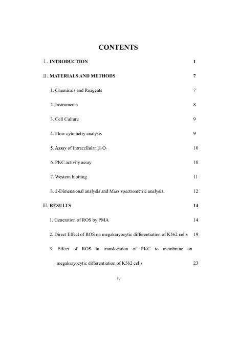
목 차
ABSTRACT 8
Ⅰ. INTRODUCTION 10
Ⅱ. MATERIALS AND METHODS 16
1. Chemicals and Reagents 16
2. Instruments 17
3. Cell Culture 18
4. Flow cytometry analysis 18
5. Assay of Intracellular H₂O₂ 19
6. PKC activity assay 19
7. Western blotting 20
8. 2-Dimensional analysis and Mass spectrometric analysis 21
Ⅲ. RESULTS 23
1. Generation of ROS by PMA 23
2. Direct Effect of ROS on megakaryocytic differentiation of K562 cells 28
3. Effect of ROS in translocation of PKC to membrane on megakaryocytic differentiation of K562 cells 32
4. Effect of ROS in activation of ERK on megakaryocytic differentiation of K562 cells 35
5. Differentiation pattern of human erythroleukemia K562 cells by glucose oxidase 38
6. Gene expression changes by ROS in megakaryocytic differentiation of K562 cells 41
Ⅳ. DISCUSSION 44
Ⅴ. REFERENCES 54
Ⅵ. 논문개요 70
Fig. 1 PMA induces the megakaryocytic differentiation of K562 cells 25
Fig. 2 Generation of ROS by PMA 26
Fig. 3 Inhibition of megakaryocytic differentiation of K562 cells by antioxidants 27
Fig. 4 H₂O₂ induces the megakaryocytic differentiation of K562 cells 30
Fig. 5 Inhibition of H₂O₂-induced megakaryocytic differentiation of K562 cells by antioxidants 31
Fig. 6 Activation of PKC by H₂O₂ on megakaryocytic differentiation of the K562 cells 33
Fig. 7 Inhibition of CD41 expression by PKC inhibitor, GF109203X, on ROS-induced megakaryocytic differentiation of the K562 cells 34
Fig. 8 Activation of ERK activity by H₂O₂ on megakaryocytic differentiation of the K562 cell line 36
Fig. 9 Inhibition of CD41 expression by ERK inhibitor, PD98059, on megakaryocytic differentiation of K562 cells 37
Fig. 10 Differentiation pattern of ROS-induced differentiation of K562 cells 40
Fig. 11 Detection of glucose oxidase-responsive protein in K562 cells 42
Fig. 12 Mass spectrometirc analysis of changed protein spots 43
Fig. 13 Scheme of megakaryocytic differentiation of K562 cells 53
*표시는 필수 입력사항입니다.
| 전화번호 |
|---|
| 기사명 | 저자명 | 페이지 | 원문 | 기사목차 |
|---|
| 번호 | 발행일자 | 권호명 | 제본정보 | 자료실 | 원문 | 신청 페이지 |
|---|
도서위치안내: / 서가번호:

우편복사 목록담기를 완료하였습니다.
*표시는 필수 입력사항입니다.
저장 되었습니다.
