
 대표어
대표어

권호기사보기
| 기사명 | 저자명 | 페이지 | 원문 | 기사목차 |
|---|
결과 내 검색
동의어 포함
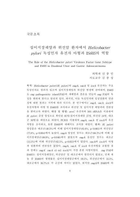
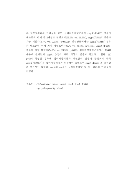
국문초록
목차
Ⅰ. 서론 9
Ⅱ. 연구 대상 및 방법 11
1. 연구 대상 11
2. 연구 방법 11
가. 내시경 검사 및 위생검 조직채취 11
나. 신속 요소분해효소검사 11
다. 세균배양검사 12
라. 중합효소연쇄반응검사 12
마. H. pylori 감염 판정 기준 15
바. 통계학적 처리 15
Ⅲ. 결과 16
1. H. pylori 양성률 16
2. cagA의 발현 16
3. vacA의 발현과 아형 분석 16
4. iceA의 발현과 아형 분석 16
5. 게놈과 PAI에서 IS605의 발현 17
6. cagA, vacA, iceA 조합과 질환 발현의 연관성 분석 17
7. cagA와 IS605 조합과 질환 발현의 연관성 분석 17
Ⅳ. 고찰 19
Ⅴ. 결론 24
참고문헌 35
ABSTRACT 42
Figure 1. Expression of ureC by PCR. 30
Figure 2. Expression of 16S rRNA by PCR. 31
Figure 3. Electrophoresis after PCR amplification of H. pylori cagA and iceA. 32
Figure 4. Electrophoresis after PCR amplification of H. pylori vacA gene. 33
Figure 5. Electrophoresis after PCR amplification of H. pylori IS605. 34
*표시는 필수 입력사항입니다.
| 전화번호 |
|---|
| 기사명 | 저자명 | 페이지 | 원문 | 기사목차 |
|---|
| 번호 | 발행일자 | 권호명 | 제본정보 | 자료실 | 원문 | 신청 페이지 |
|---|
도서위치안내: / 서가번호:

우편복사 목록담기를 완료하였습니다.
*표시는 필수 입력사항입니다.
저장 되었습니다.
